

Alzheimer’s: A brain ‘avatar’ for personalised treatment based on a blood sample
Patient-derived organoids, created from the patients’ own stem cells, are currently being studied. They provide a ‘micro-replica’ of brain cells, helping us to understand how effective treatments might be and how they work. But we are only just getting started.

Key points
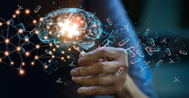
Identifying new treatments for Alzheimer’s disease is undoubtedly one of the major challenges facing neurology. But whilst targeted drugs for this condition are coming to the fore, there is another aspect to consider: given the varied ways in which the condition presents itself at its different stages, it is also essential to consider the appropriateness of treatment. In other words: each patient must be offered what they actually need at a particular stage in the progression of the disease. It is in this context that research by experts at Johns Hopkins Medicine, published in “Alzheimer’s & Dementia: The Journal of the Alzheimer’s Association” shines a spotlight on the issue. By studying a sort of microscopic ‘copy’ of the brain of those experiencing the symptoms and signs of this ‘brain fog’, it may be possible to predict patients’ responses to medication in advance. The study examined psychiatric symptoms and the effects of treatments using “organoids” – clusters of cells grown in the laboratory that have thus become miniature brains in their own right.
Real brains, scaled down
The experts, led by Vasiliki Machairaki, analysed miniature models of the rhombencephalon, an area at the back of the brain that helps regulate breathing, sleep and heart rate. The analysis focused on a drug belonging to the class of selective serotonin reuptake inhibitors, used to manage symptoms associated with the neurological condition. It all began with the collection of blood samples, from which induced pluripotent stem cells were derived – cells capable of differentiating into any cell in the body. Organoids were then created – small rhombencephalic structures containing neurons from both patients with Alzheimer’s disease and healthy individuals, capable of producing the neurotransmitter serotonin. This resulted in the creation of a miniature brain organ, about the size of a pea, and led to the study of hundreds of organoids from healthy individuals and patients.
Different responses to medication
The organoids developed using nerve cells derived from patients have, to some extent, reproduced biological characteristics typical of the disease, albeit on a molecular scale. In particular, in these ‘avatars’ of brains affected by neurodegeneration, differences were observed in the proteins involved in communication between brain cells, in inflammation and in pathways associated with the disease. Not only that: when these ‘micro-brains’ were treated directly with a drug widely used in clinical practice (escitalopram), different responses to the same treatment were observed. Specifically, in some organoids derived from patients, the treatment did indeed increase – as intended – the levels of proteins involved in serotonin signalling and communication between brain cells. However, other organoids showed little or no molecular response. “We used these organoids to model how tissue from certain patients might respond to a commonly prescribed SSRI,” commented Machairaki in a university press release. “On a larger scale, our model could be used in the future to identify subgroups of patients, based on underlying molecular mechanisms, who are more likely to respond to certain drugs, thereby helping us to develop precise, targeted long-term treatments.”
Possible biomarkers of the condition
As if that weren’t enough, the research reveals another interesting aspect. The researchers also analysed specific extracellular vesicles released by the organoids, to assess their potential usefulness as biomarkers for Alzheimer’s disease or as a means of understanding the tissue response, by evaluating the proteins within the extracellular vesicles themselves before and after treatment of the organoids with escitalopram, the proteins within the extracellular vesicles themselves. It was found that these tiny, invisible vesicles contain proteins involved in essential brain functions, including communication between neurons, memory and the release of neurotransmitters. In short: they could potentially prove to be not only indicators for monitoring the condition, but also markers of response to treatment.
Fascinating but not immediate prospects
“The study opens up a range of possibilities for a future that is certainly not imminent, primarily because it captures the potential genetic and biological variability of individual patients and thus enables tissue typing from the patient’s blood – comments Vincenzo Andreone, director of the Complex Operational Unit of Neurology and Stroke Unit at Cardarelli Hospital in Naples –. However, we are only talking about psychiatric symptoms here, so not about therapies that can alter the course of the disease, but treatments for disorders such as insomnia, agitation, depression and hallucinations, which affect around 90 per cent of patients. Research suggests that the in vitro organoid, as a research model, can capture different responses to the drug, and it will take a long time.”

