

Arrhythmias and more: when and how does the ‘electrical system’ change following cardiac surgery?
Sometimes surgery can trigger arrhythmias, making it necessary to have a pacemaker or defibrillator fitted: what to do and how to monitor the situation

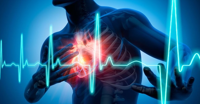
Imagine a city where electrical signals run through fibres beneath the tarmac. At some point, in a particular neighbourhood, the lights go out – perhaps because there’s a fault in one of the power lines. Or they flicker on and off beyond anyone’s control. Something similar can happen in the heart too. The electrical signal that coordinates the regular flow of contractions between the atria and ventricles can become disrupted. To understand what can happen, let’s take a look at the heart’s electrical ‘pathways’. The starting point, the heart’s natural pacemaker, is the sinus node, which is located in the right atrium and generates the electrical impulse: this first spreads through the atria and then travels down to the atrioventricular node, where the signal is ‘routed’ to the ventricles via anatomically defined pathways. The pathway continues through the bundle of His, which originates from the atrioventricular node, travels downwards and divides into a left and a right branch before descending to the lower part of the ventricles. Although cardiac surgery is becoming increasingly minimally invasive, the scalpel can also trigger an irregular heart rhythm or exacerbate its severity. For this reason, both before and after the operation, the role of a cardiologist specialising in electrophysiology is crucial, working as part of the team caring for the patient.
A study warns
It is extremely important to pay attention to heart rhythm disorders. Whilst, on the one hand, it is sometimes necessary to implant a pacemaker to regulate the heart rhythm or a defibrillator to deliver a ‘shock’ in the event of ventricular fibrillation – thereby restoring the ventricle’s electrical activity to normal and preventing the risks associated with the heart’s inability to pump blood effectively – on the other hand, it must be borne in mind that the presence of these devices may be associated with an increased risk of hospital admissions for heart failure or death. This is highlighted by a study published in *Heart*, coordinated by Laura Alice David of the University of Gothenburg. The study, which does not establish a cause-and-effect relationship but merely reveals an association, examined the long-term outcomes of the implantation of a permanent pacemaker or a defibrillator shortly after cardiac surgery. Over the course of nearly seven years of follow-up, data from Swedish registers on over 76,000 people were analysed. Of these, 2.8 per cent had a permanent pacemaker fitted and 1 in 200 had an implantable defibrillator fitted within a month of the operation. The result: risks increase in the presence of such devices. The authors themselves note that pacemaker implantation is often unavoidable and life-saving. However, focusing on pacing strategies for these patients that reduce the risk of the ventricles contracting out of synchrony, and paying close attention to any early signs of heart failure, could prove helpful in the future.
What to do and who is most at risk
“Certainly, in patients undergoing cardiac valve surgery – particularly when it comes to the aortic valve – there is a risk of impaired conduction at the atrioventricular node, due to mechanical compression, ischaemia or tissue damage,” comments Giulio Molon, Director of the Complex Operative Unit of Cardiology at the IRCCS Sacro Cuore Don Calabria in Negrar (Verona). “In such cases, pacemaker stimulation is required.” However, caution is required: the traditional method of apical right ventricular pacing could, however, cause intra- and interventricular contraction dyssynchrony, potentially leading to cardiomyopathy, heart failure and atrial fibrillation, as reported in the study. “To avoid this problem, ventricular catheters – and more recently atrial catheters too – have been implanted in the conduction system for several years now,” notes Molon, who also points out that particular attention must be paid to patients undergoing cardiac surgery such as coronary artery bypass grafting.”. In such cases, it is more common to find a history of a previous heart attack with cardiac scarring – sometimes extensive – and even severe reductions in ventricular contractility. For this reason, too, it is not uncommon to encounter cases of tachycardia or ventricular fibrillation, and a defibrillator may therefore be implanted to prevent sudden arrhythmic death; however, above all, attention must be paid to the risk of progression to heart failure.
Remote monitoring
“A highly effective approach in these patients for reducing such events is management via telemedicine – that is, the use of transmitters or apps that allow the patient’s pacemaker or defibrillator to be connected directly from their home to the implant centre,” concludes the expert. In this way, we are able to detect early warning signs of heart failure well in advance, even before the patient experiences any symptoms, enabling us to initiate treatment if necessary, carry out outpatient assessments or take other measures to prevent overt, clinical heart failure. ‘We have been monitoring all our patients with implanted devices since 2008, and this has drastically reduced the number of emergency admissions among these patients.’
In particular, remote management allows for remote monitoring of devices: the specialist receives data from the device and, even before symptoms of heart failure appear, can consult with the patient, advise them on adjustments to their treatment and/or changes to their lifestyle ordiet, as well as advising on a possible consultation or admission to fine-tune treatments before an emergency hospital admission due to heart failure, which worsens the patient’s prognosis.

