
Interventional cardiology

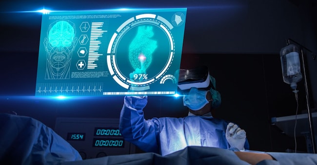
Heart to repair, angioplasty 3.0 at Gemelli is done with AI

A 'new era' for diagnosis and therapy: the Roman polyclinic is the first hospital in Italia to use new artificial intelligence software to guide coronary treatment

by Health Review

 Adobe Stock

5' min read

Translated by AI
Versione italiana

5' min read

Translated by AI
Versione italiana

At the Policlinico Universitario A. Gemelli Irccs - first in Italy - artificial intelligence is coming to support interventional cardiology 3.o. That is, technology - the hospital explains - that is increasingly precise, personalised and oriented towards clinical results. So much so as to open 'a new era for the precision of diagnosis and therapy'.

Angioplasty 3.0

The 'upgrading' is entrusted to a new software with AI to guide coronary angioplasty: the Rome-based Irccs is already one of the main centres of reference for interventional cardiology in Italia and related research, and thanks to the clinical and scientific experience of its specialists, it has been chosen as the first hospital in Italia and one of the first three centres in the Emea area (Europe, Middle East and Africa), for the introduction of Ultreon 3.0, an innovative software based on artificial intelligence that makes it possible to make coronary revascularisation (stent) procedures guided by endocoronary imaging even more precise and personalised. The official debut of the new technology was on 30 March, in the Gemelli's haemodynamics room, with the first clinical use marking the entry of a new generation of instruments for interventional cardiology.

Loading...

The procedure

The pivotal examination for identifying the lesions responsible for a heart attack and planning its treatment has been coronarography or coronary angiography for decades. This procedure makes it possible to visualise the coronary arteries - the vessels that nourish the heart - by injecting an X-ray contrast medium into them. To perform it, the cardiologist introduces a special catheter into an arterial vessel - nowadays increasingly from the radial artery of the wrist - and guides it to the origin of the coronary arteries, in the root of the aorta. From here, the contrast medium allows any narrowing or occlusions due to atherosclerotic plaques to be detected; based on this, the cardiologist plans the revascularisation treatment.

L’evoluzione

In recent years, this technique has evolved significantly thanks to the integration with intracoronary imaging systems, such as Oct (Optical Coherence Tomography), which is currently the method that allows us to observe plaque from inside the vessel with the highest level of detail.

In this context, Policlinico Gemelli stands out as one of Italy's leading centres for the use of Oct, on the strength of its long clinical experience and significant scientific production in the field.

The technology

What makes this diagnostic technology even more advanced and precise today is the introduction of new software that received the CE mark on 11 March. This is the first system available in Europe that can use Oct images to assess both the pressure drop along the vessel and the structural characteristics of the obstruction, thus concretely supporting the clinician in the choice and optimal positioning of the stent, with the aim of maximising the improvement of blood flow.

The solution incorporates a one-second Oct pullback, providing higher resolution images than intravascular ultrasound (Ivus). It also significantly reduces - up to zero in some cases - the use of contrast medium during the procedure and introduces advanced predictive capabilities and artificial intelligence-based insights for even more accurate lesion assessment.

A new era

"Coronarography and angioplasty have historically been based on angiographic images, which, however, have a limited resolution capacity," explains Professor Francesco Burzotta, Professor of Cardiology at the Catholic University of the Sacred Heart and Director of the Uoc of Cardiology at Policlinico Gemelli. To improve the accuracy of therapeutic decisions we now use two complementary approaches: the functional study of the coronary arteries through Fractional Flow Reserve (Ffr), which measures how much a plaque obstructs blood flow, and intracoronary imaging techniques such as Oct, which allow us to visualise the artery from the inside'. Gemelli has been an international leader in the use of Ffr and Oct for years, both clinically and scientifically, with numerous publications that have contributed to defining the most advanced strategies in the management of ischaemic heart disease.

AI at the service of the cardiologist

But it can always be improved. The new software, a next-generation, artificial intelligence (AI)-based Oct imaging platform capable of providing doctors with usable clinical information in real time, is a further step forward. Thanks to artificial intelligence, the system automatically analyses Oct images, allowing the potential functional impact to be estimated. 'The real novelty,' explains Professor Carlo Trani, Professor of Cardiology at the Università Cattolica del Sacro Cuore and Director of the Uoc Interventional Cardiology and Invasive Diagnostics of Fondazione Policlinico Gemelli, 'is that from the Oct image the software is also able to derive functional information similar to that of the Ffr. Traditionally, these two pieces of information required the use of two different catheters, with an increase in procedural time and costs. The new technology, on the other hand, allows the two assessments to be integrated from a single Oct acquisition, facilitating faster and more personalised therapeutic decisions'.

'We don't think this innovation will completely replace Fractional Flow Reserve, whose validity is supported by numerous clinical studies,' Burzotta points out, 'but it certainly represents a long-awaited development that opens up a new phase in the use of intracoronary imaging.

The impact on therapy

The information obtained thanks to the new software also has a direct impact on the therapeutic phase. "When we perform an angioplasty and place a stent - a small metal prosthesis that keeps the artery open - we have to make sure that it expands perfectly and in the correct position," Trani emphasises. "Oct's advanced analysis allows us to predict and optimise the outcome, improving the accuracy of the procedure.

"As cardiologists continue to adopt Oct and move away from traditional imaging methods such as angiography, emerging technologies become critical in defining the best course of care for the patient. Thanks to artificial intelligence, the software is able to automatically detect vessel calcium and diameter, integrating imaging and physiology information into a single platform and allowing physicians to place stents exactly where they are needed," - comments Evelina Cerri, General Manager of Abbott's vascular division in Italia.

Ischaemic heart disease, often caused by the development of atherosclerotic plaques in the coronary arteries, remains a major cause of mortality. Understanding the biological structure of the plaque and its impact on blood flow is crucial for deciding when and how to intervene. Oct allows the plaque to be studied with a level of detail comparable to in vivo microscopy, while functional assessments indicate how much the narrowing compromises oxygen supply to the heart, especially during exertion. 'The possibility of obtaining both of these pieces of information in a few seconds,' Burzotta concludes, 'allows us to make even more precise and personalised therapeutic decisions, with the aim of improving the outcomes of revascularisation procedures and reducing the risk of cardiovascular events.

Copyright reserved ©
Loading...

Brand connect

Loading...

Newsletter

Notizie e approfondimenti sugli avvenimenti politici, economici e finanziari.

Iscriviti