

Precision oncology, an 'open' AI tool from Microsoft Research to study tumours and identify the best treatments
Presented in the journal Cell the first population-scale study that maps the tumour immune microenvironment using virtual spatial proteomics: a technology analyses and interprets the data with a view to targeted interventions
Key points
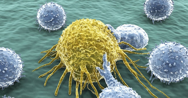
"GigaTime is proof of what is possible when cutting-edge artificial intelligence meets real clinical data at scale. Working closely with Providence and the University of Washington, we have demonstrated how multimodal AI can transform routine anatomical pathology slides into rich spatial proteomic maps, making once unattainable discoveries possible. Our hope is that, by making GigaTIME freely accessible, we can accelerate research and help the entire field move towards more precise and personalised cancer treatments'. This is how Hoifung Poon, General Manager of Real-World Evidence at Microsoft, outlines the prospects of the new AI tool in the service of the fight against cancer, developed by Microsoft Research. It is the first population-scale study to map the tumour immune microenvironment using virtual spatial proteomics, an AI technology that analyses and interprets data. This makes it possible to identify previously invisible patterns and relationships, including new links between genetic mutations and protein activations.
The Studio
Presented in a paper in the scientific journal Cell - GigaTime allows researchers to study the tumour microenvironment on a previously unseen scale, a key element in predicting how tumours behave and which therapies work best. In the article, the researchers summarise the scope of the innovation as follows: The tumour immune microenvironment (Time) has a critical impact on cancer progression and immunotherapeutic response. Time' is in fact a highly complex spatial ecosystem consisting of tumour cells and several non-malignant cell types, including immune cells, cancer-associated fibroblasts (Caf), endothelial cells (Ec), pericytes and other cell types, embedded in an altered extracellular matrix. Multiplex immunofluorescence (mIF) is an excellent tool for multichannel protein profiling co-localised on the same tissue, preserving the spatial architecture, but its use remains limited by the substantial cost for large-scale study due to reagents, specialised equipment and computational infrastructure, combined with labour-intensive workflows for staining, imaging and data processing. As a result, existing mIF datasets are extremely scarce, which significantly limits their applicability in clinical discovery and translation.
In contrast, haematoxylin and eosin (H&E) images are routinely generated in low-cost clinical workflows for the study of tissue structure and cell morphology. And if an H&E image does not explicitly reveal cell states, the spatial configuration of the cells it highlights can shed light on their individual states. These patterns, the researchers warn, may not be obvious to human eyes but are potentially discernible using advanced multimodal AI. Not only that, the latest advances in AI further amplify this potential, as AI demonstrated superior performance with pre-training on a large collection of pathology images.
The Instrument
The tool, Microsoft emphasises, builds on Microsoft's ongoing work in advancing multimodal GenAI to scale the generation of Real World Evidence. Projects such as GigaPath, BiomedParse, Curiosity, and Trialscope aim to develop 'virtual patients' i.e. AI-based models that predict health outcomes and guide personalised care decisions.
Based on models capable of processing multiple types of data, GigaTime could help change the way researchers study cancer, bypassing the problem of expensive and time-consuming laboratory tests to understand how tumours develop in the body. Instead, the AI tool - Microsoft Research points out - transforms 'common, low-cost pathology slides into complex and detailed digital maps showing how immune cells interact with cancerous tumours through protein activation, enabling researchers to uncover previously invisible patterns'. This opens up new opportunities to study tumour microenvironments on an unprecedented scale. Generating these kinds of digital maps would take days and thousands for a single sample, whereas with AI it is possible to simulate these analyses on dozens of proteins in seconds thanks to computational processing, enabling the study of thousands of scenarios simultaneously.

